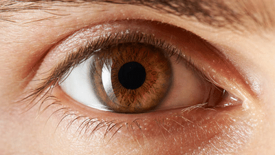
A patient walks into an ophthalmologist's office worried about blurry vision. What the doctor sees during that exam might actually say more about the patient's heart than their eyesight. That's not a coincidence, and it's not new either, though the science backing it up has gotten a lot sharper recently.
Blood vessels aren't usually something a doctor can just look at directly. Cutting someone open is generally how you'd see an artery up close. The retina breaks that rule entirely. It's the one place in the body where a physician can photograph actual blood vessels in seconds, no incision required, just a light and a camera pointed at the back of the eye. Those tiny vessels running across the retina's surface happen to mirror what's going on elsewhere in a person's circulatory system, including the vessels feeding the heart, which are nowhere near as easy to examine directly.
Doctors have suspected this link for decades, honestly. What's actually shifted is how precisely it can now be measured. A prospective multi-center clinical trial presented at the American College of Cardiology's 2026 scientific sessions found that an AI system reading retinal images could flag people at elevated cardiovascular risk with 91.1 percent sensitivity and 86.2 percent specificity, numbers that held up well against standard risk assessments built around blood pressure and cholesterol testing. Around a quarter of the participants in that trial came back with elevated cardiovascular risk using conventional methods. The retinal scan picked up almost the exact same group, and did it with nothing more than a photograph.
What this actually means for patients is fairly significant. Narrowing vessels, odd branching patterns, tiny areas of bleeding visible in a retinal scan can signal hypertension or diabetes related vascular damage years before someone develops symptoms serious enough to book a cardiology appointment.
This is exactly why routine eye screening deserves a second look, particularly for anyone managing diabetes or high blood pressure, since vascular damage in these conditions tends to progress without much warning. At Sharp Sight Eye Hospitals, this overlap between eye health and heart health has shaped how diabetic retinopathy screening runs across the network. Combining advanced retinal imaging with AI assisted diagnostics lets clinicians spot microvascular changes early enough that intervention still has real impact, instead of catching the problem after vision has already started slipping.
For most readers, the takeaway is pretty simple even though the underlying science isn't. Getting a dilated eye exam, especially with diabetes, high blood pressure, or a family history of heart disease, does more than protect eyesight. It offers a genuine look at vascular health that few other non-invasive tests can match. Skipping it just because vision feels fine misses the whole point, since damage to retinal vessels usually shows up well before any change in eyesight does. Waiting for symptoms before getting checked tends to backfire for exactly that reason.
None of this replaces an actual cardiology workup, and nobody should treat an eye exam as a substitute for a blood pressure check or a lipid panel. But as a cheap, non-invasive layer that catches people who might otherwise go years without realising their vascular risk is climbing, this connection between eye and heart deserves a lot more attention in routine checkups than it currently gets. With diabetes and cardiovascular disease both on the rise across the country, that five minute retinal scan might be doing more preventive work than most people give it credit for.
Dr. Vipul Singh, Senior Consultant, Sharp Sight Eye Hospitals
Contact to : xlf550402@gmail.com
Copyright © boyuanhulian 2020 - 2023. All Right Reserved.